

Inflammatory Bowel Disease (IBD) Models
Inflammatory bowel disease (IBD) is a chronic nonspecific inflammatory disease. It mainly includes Crohn disease (CD) and ulcerative colitis (UC).

1. DSS-induced UC model
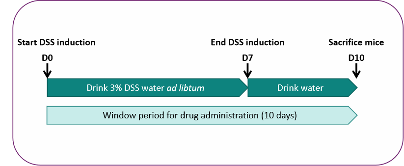
Validation data
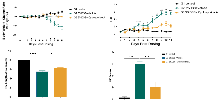
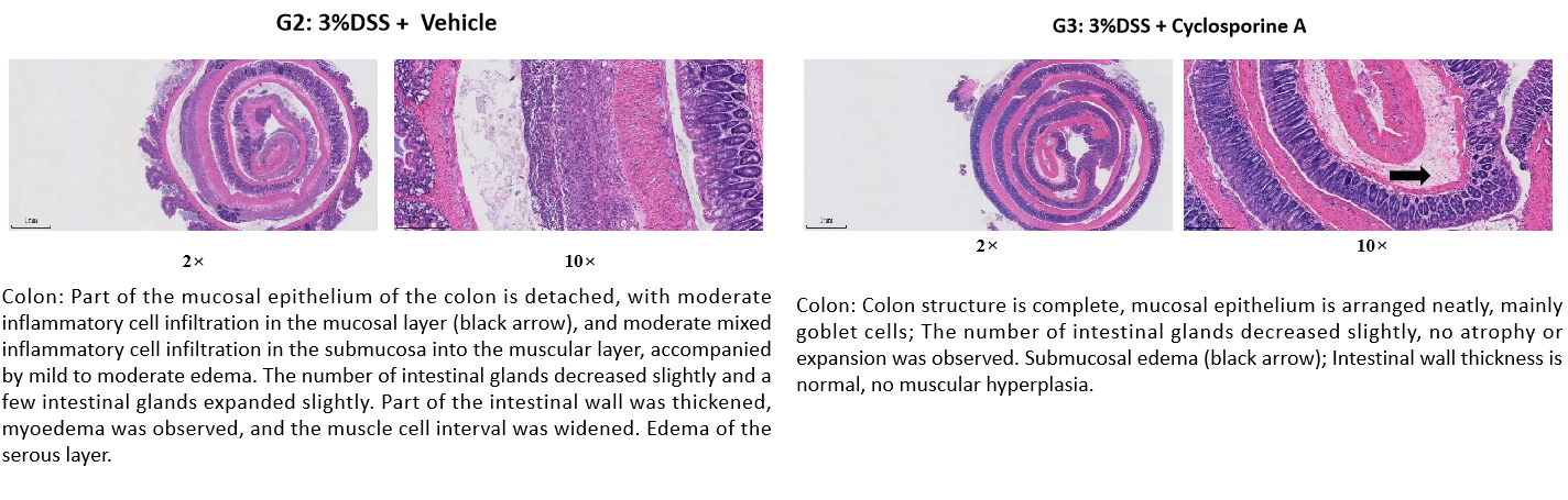
Cyclosporine A (CsA) alleviated weight loss and decreased disease activity index (DAI) score in UC mice. And the pathological symptoms such as epithelial exfoliation of colon mucosa and inflammatory cell infiltration were relieved.
2. DNBS-induced colitis model
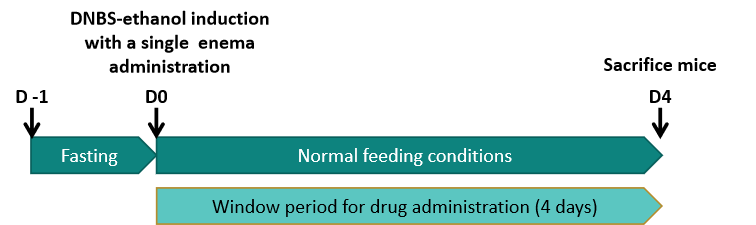
Validation data
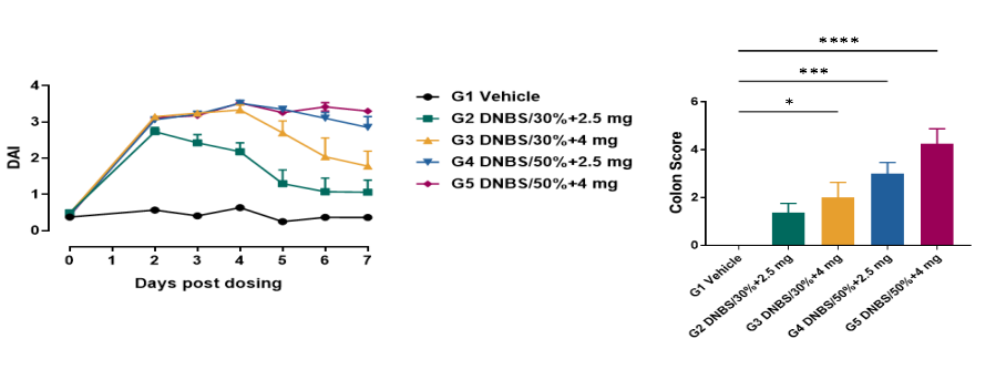
Compared with G1 group, DAI score in model group was significantly higher.
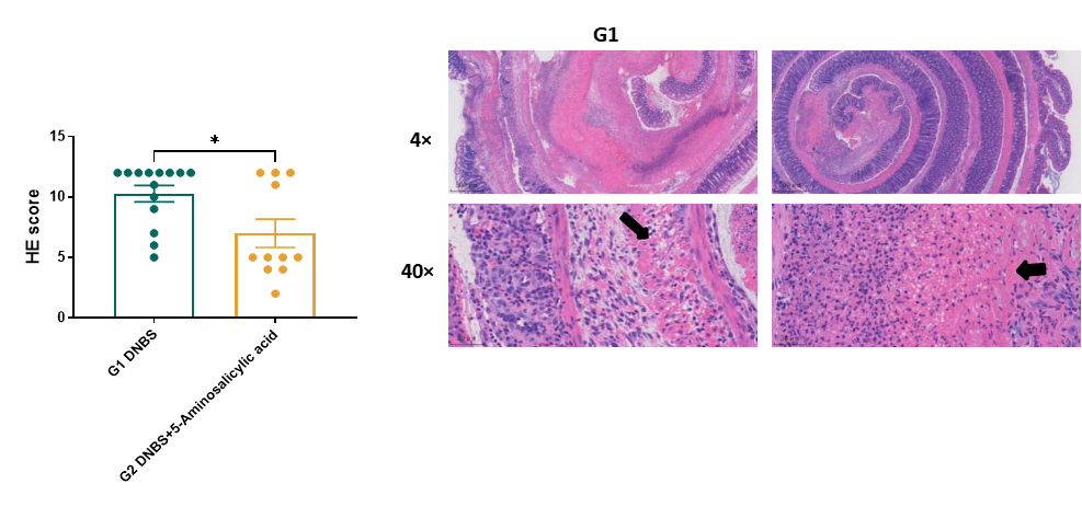
After treatment with 5-aminosalicylic acid in G2 group, HE pathological score decreased significantly, and pathological symptoms such as necrosis, exfoliation and inflammatory cell infiltration of colonic mucosa were relieved
3. IL-10 KO colitis model
Model features of B6-IL-10 KO mice

Chronic IBD model that is capable of characterising more accurate histological phenotypes than actue IBD models;
Spontaneously aqucire colitis-related colonic inflammation after 6 months;
Extented and abundant window period for drug administration. More suitable for the investigation of probiotics and prebiotics;
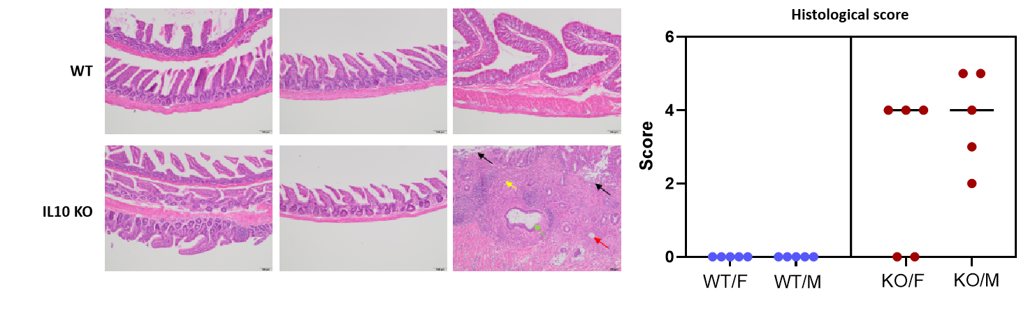
No obvious pathological changes were observed in jejunum and ileum; Degeneration/necrosis/deletion of the mucosal epithelium and intestinal glands were observed in colon; Inflammatory cell infiltration and fibrous tissue hyperplasia in Lamina propria, submucosa, muscularis, adventitia; Few muscular cyst was observed; The incidence and severity of male were slightly higher than those in females.
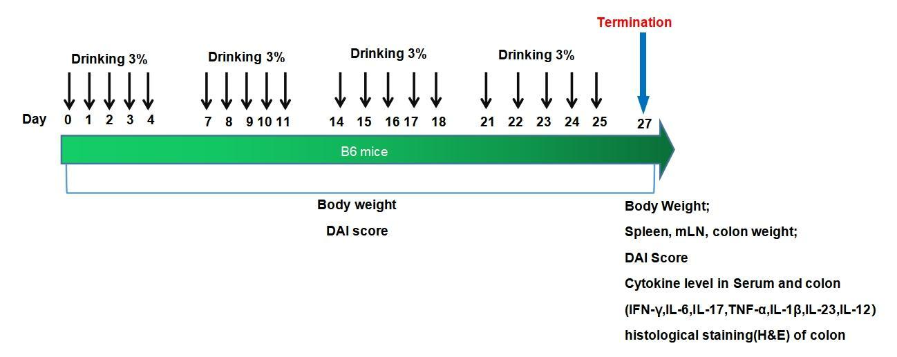
4. Dextran sodium sulfate (DSS)-induced chronic colitis model
Experimental mouse strains: B6, 6-7weeks old, male
Modeling cell: 3% DSS
Modeling method:
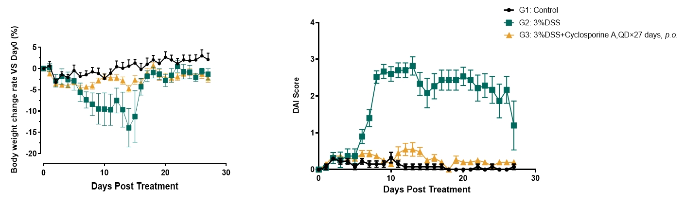
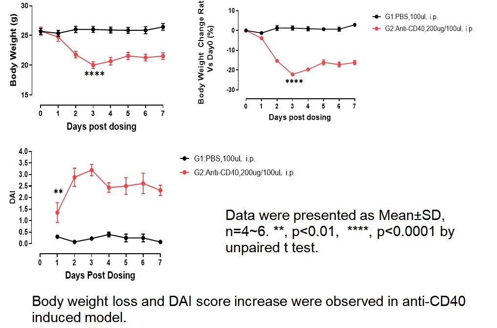
Body weight-loss and DAI score increase were observed in DSS induced model.
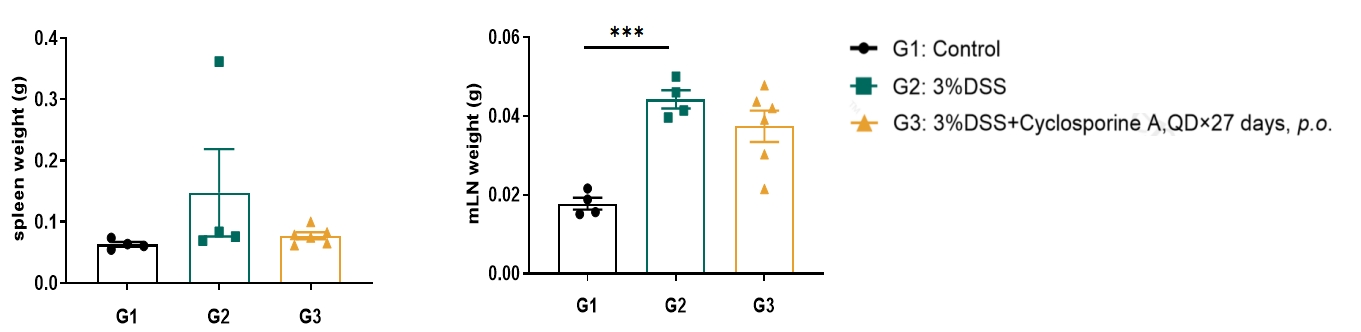
Mesenteric lymph node (mLN) weight increase was observed in DSS induced model.
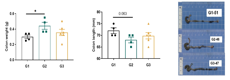
Colon weight increase and length decrease were observed in DSS induced model.
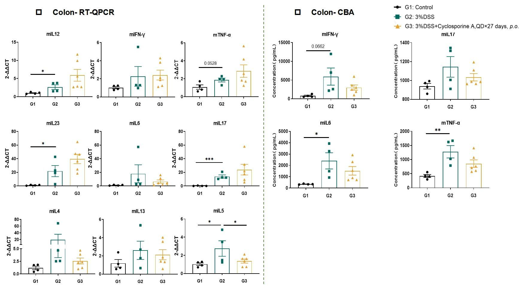
Th1/Th2/Th17 cell involved in DSS induced chronic colitis model,especially Th17 cell.

5. Anti-CD40 induced colitis model
Model strategy
Experimental mouse strains: RAG-/-, 6-7weeks old, female
Modeling reagents: anti-CD40 antibody
Modeling method:
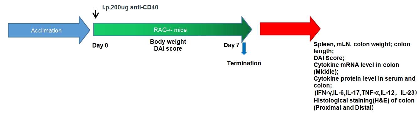
Short modeling time (7 days), ease of use (upon injection of anti-CD40 antibody);
Evaluate the innate immune response in disease using cytokine profiling of : IFN-γ、IL-12、IL-23、TNF-α.
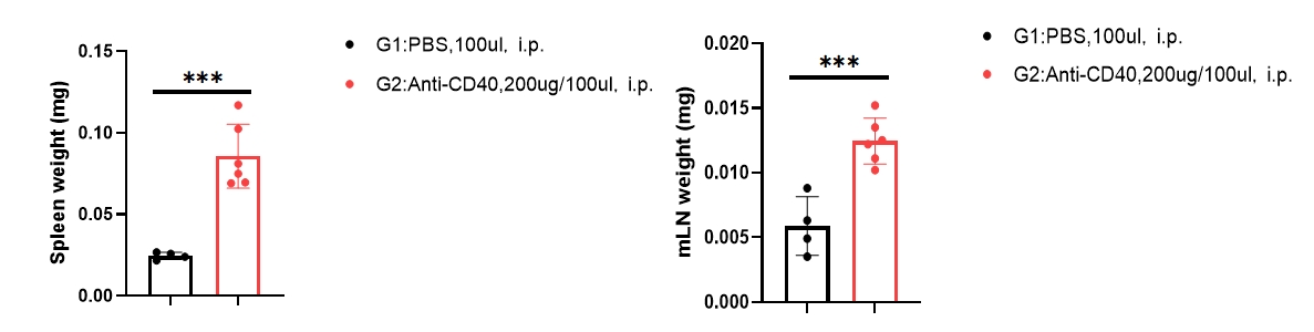
Spleen and mesenteric lymph node(mLN) weight increase was observed in anti-CD40 induced model.
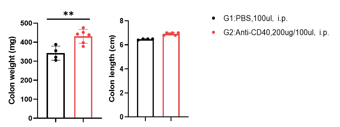
Spleen, mesenteric lymph node (mLN) and colon weight increase was observed in anti-CD40 induced model.
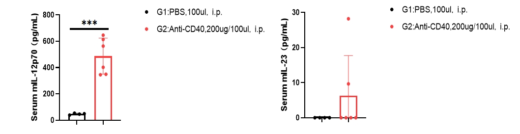
ELISA
Cytometric bead array
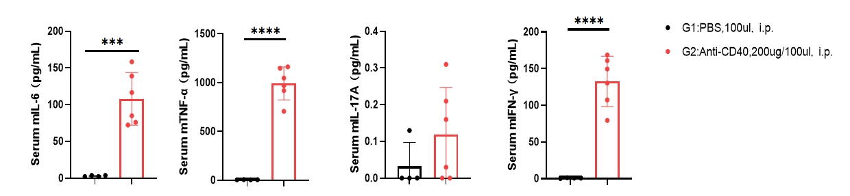
Serum IL-12, IL-23, IL-6,TFN-α and IFN-γ levels were elevated in anti-CD40-induced model.
Cytometric bead array
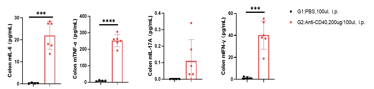
Colon IL-6,TFN-α and IFN-γ cytokines protein level was significantly increased in anti-CD40-induced model.
RT-QPCR
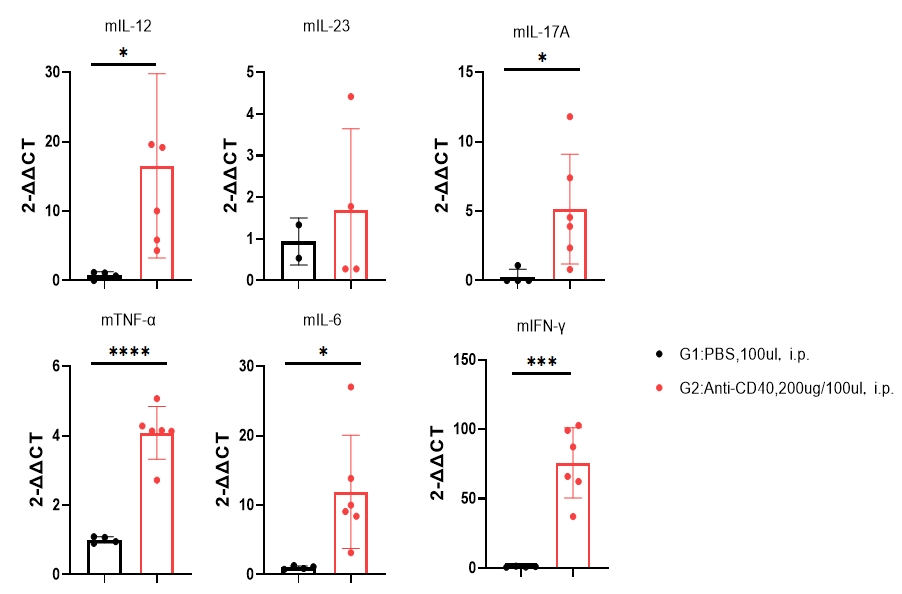
Colon IL-12, IL-6,TFN-α, IL17A and IFN-γ cytokines mRNA level was significantly increased in anti-CD40 induced model.
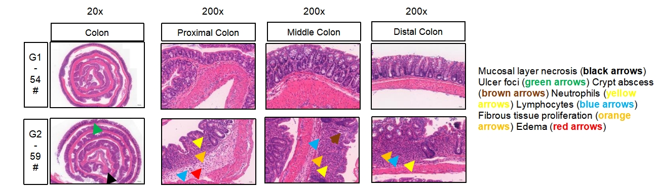
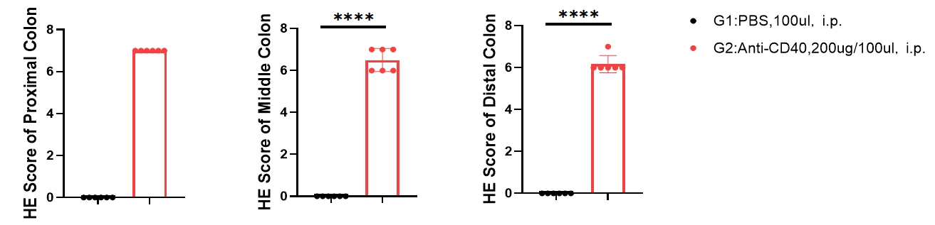
H&E staining of colon demonstrating histopathological features associated with anti-CD40 induced colitis