

Alopecia Areata (AA)
Alopecia areata (AA) is a common chronic tissue-specific autoimmune disease that leads to hair loss and affects up to 2% of the general population. It is characterized by unpredictable episodes of hair loss that relapse and remit, impacting the scalp, facial hair, eyebrows, eyelashes, and body hair. While AA does not typically affect physical health, it can have significant psychosocial and psychological effects. The exact cause remains unknown, but genetics, stress, and environmental factors may contribute. Disease onset of AA is hypothesized to follow the collapse of immune privilege of the hair follicle, which results in an increase in self-peptide/MHC expression along the follicular epithelium. Hair loss is associated with infiltration of the hair follicle with putatively self-reactive T cells. Interferon-γ (IFNγ) has been identified as the key player in AA pathogenesis.
Skin‐draining LN cells graft C3H/HeJ mouse
C3H/HeJ mice is the most widely used murine AA model that has long dominated preclinical AA in vivo research. Aging C3H/HeJ mice can develop a spontaneous, complex polygenic, AA-like hair loss that undergoes stages of waxing and waning. However, the spontaneous incidence of AA in C3H/HeJ mice is relatively low, with only 0.25% at 5 months and 20% at 18 months.
To establish a highly reproducible AA model, Here we utilize lymph node cells(LNCs) from AA-affected mice and graft these LNCs to young normal-haired C3H mice. This approach provide a highly reproducible model with progressive lesions that makes it be a perfect AA model for drug efficacy and mechanism-based studies.
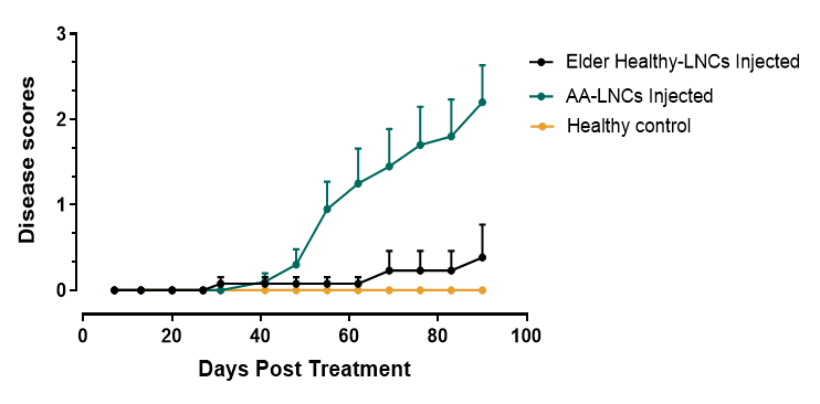
Fig.1 AA-disease score of LNCs induced C3H mouse
Gross lesions were scored subjectively on a scale of 0-6 (ref PMID 39105684). Gross photographs of ventral skin and dorsal skin were taken every week from the start until completion (time of euthanasia) of the study. Grade 0: Mice had completely normal hair length and distribution on all surfaces of the body. Grade 1: Faint evidence of hair loss, particularly in the axillary, inguinal, and distal mammary gland regions.Grade 2: Obvious patches of hair loss, particularly on the ventral body surface.Grade 3: Large areas of obvious hair loss but also remnants of areas of normal haired areas.Grade 4: Large areas of hair loss with small remnants of areas of normal hair.Grade 5: Almost completely bald on the ventral surface and large areas of hair loss on the dorsal surface. Grade 6: Total alopecia on the ventral and dorsal surface.
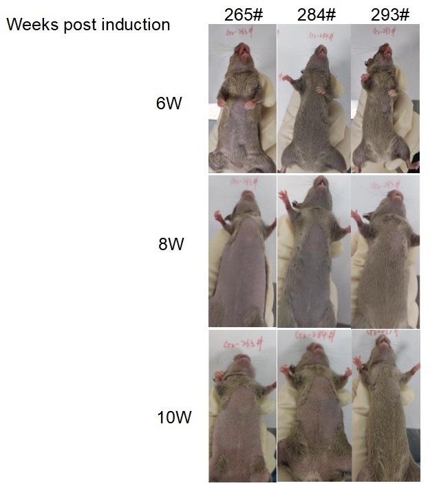
Fig.2 Progress of AA-hair loss over time